.avif)
Vireo™
The world's fastest live-cell imaging system
From high-throughput discovery to uncovering modes of action, Vireo™ accelerates your research with real-time live-cell imaging.


01
Unmatched Throughput
1-minute per plate means more experiments and less waiting
Process 1,000 plates per day and 1,000 conditions per plate with Vireo’s high-speed imaging. Optimized for brightfield and multi-channel fluorescence, its automated z-stacking ensures rapid data acquisition while preserving sample integrity and preventing photodamage to living cells.
Video Link
02
Automation-Ready
Compatible with robotic systems for automated workflows
Vireo™ integrates seamlessly with lab automation platforms to enhance throughput and scalability. Our content-aware focusing and segmentation pipelines simplify workflows, enabling teams to achieve reproducible results with minimal intervention, saving time and eliminating tedious image processing steps.
Video Link
03
Parallelized Video
Function and dynamics at scale
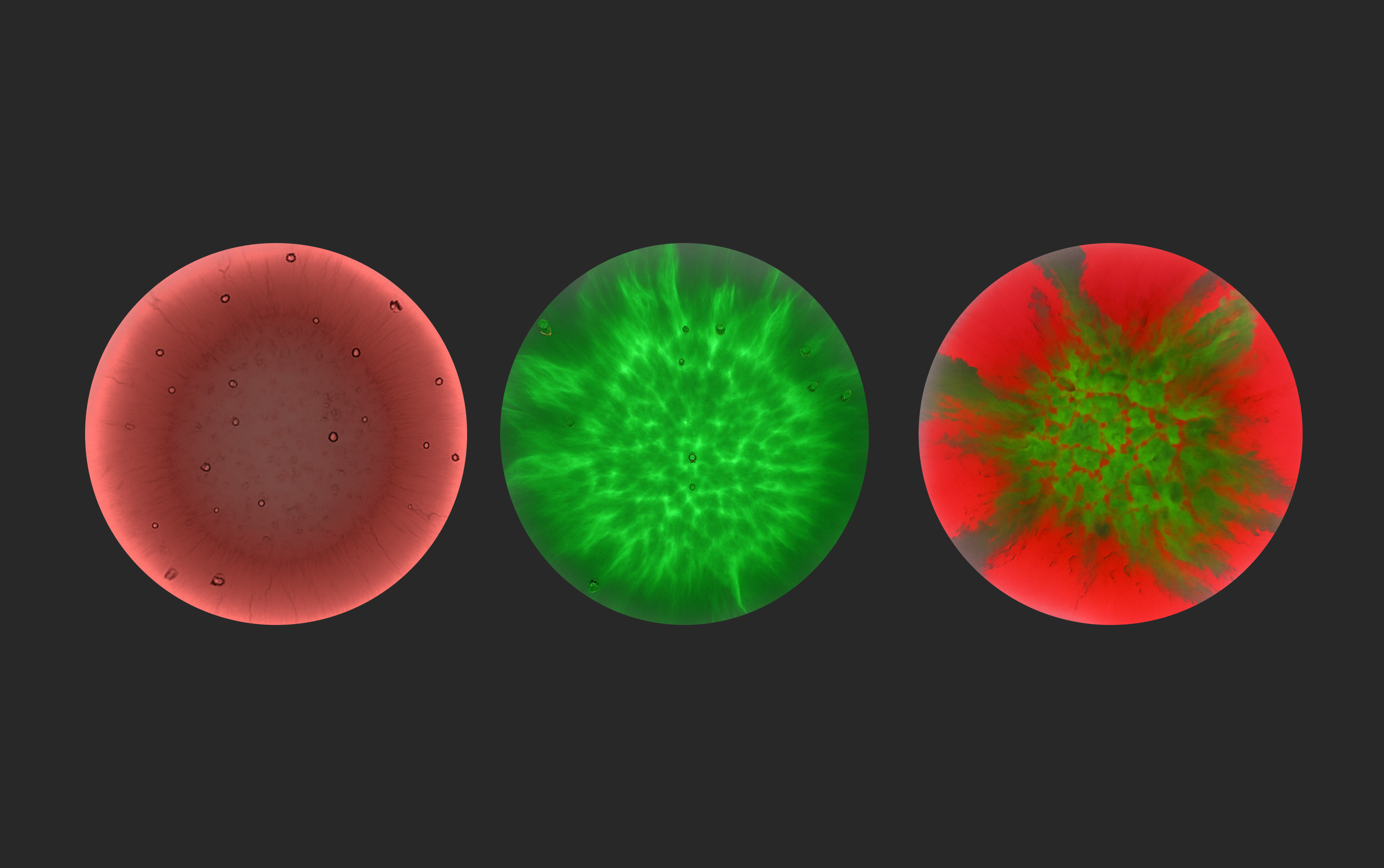
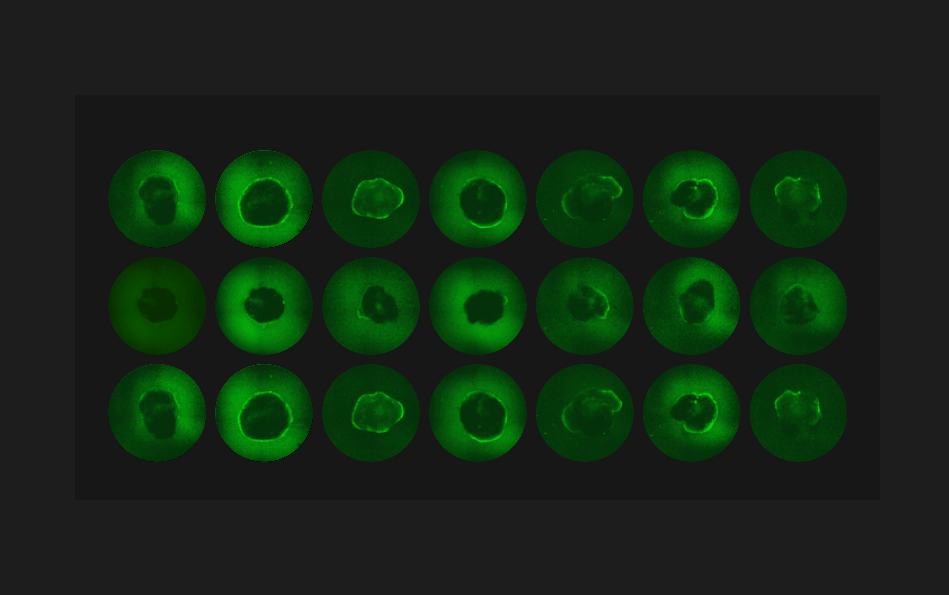
Analyze multi-dimensional video microscopy data across 24x experimental conditions synchronously for unbiased and non-destructive assessments of cellular function and dynamics. Moving from longitudinal to continuous observation delivers rich data about sub-populations, internalization speeds, and physiological processes - at scale for the first time ever.
Video LinkBrightfield imaging at 0.5um/px
Single-cell resolution Automated z-stacking
Single-cell resolution Automated z-stacking
.avif)
Get Vireo™ up and running with ease
.avif)
.avif)
MCAM™ software is exclusively designed for use with Ramona’s Kestrel™ and Vireo™ microscope systems.
Contact our sales team to explore pricing packages tailored to your research needs.
.avif)
Quick Setup
Designed for plug-and-play simplicity, Vireo™ can be up and running quickly with minimal disruption. Streamlined hardware and intuitive software ensure researchers can focus on discovery, not troubleshooting.
Robot-ready
Compatible with scheduling software through a formal Web API, Vireo™ integrates effortlessly into automated workflows for maximum throughput.
Built for Daily Use
Vireo™ combines the power of a room full of microscopes into a compact benchtop system designed to get researchers the data they need 24x faster so they can stay focused on the biology.
01
Optical Characteristics
Field-of-view
behavior mode
SBS multi-well plates (120mm x 80mm)
screening mode
SBS multi-well plates (120mm x 80mm)
Array Size
24x video microscopes
24x video microscopes
Simultaneous video
Full plate
Partial plate
Resolution
9.1 µm/px
3.1 µm/px
Working Distance
240 mm
90 mm
Depth-of-field
3000 µm
500 µm
02
Sensor Characteristics
Image Sensors
CMOS - Color
Array Pixel Count
315 Megapixel
Bit Depth
8
Dark Current (Typ.)
5.9 LSB/s
SNR (Typ.)
35 dB
Peak QE
0.92
Digital Gain
7
Analog Gain
2
Minimum Exposure
150 microseconds
Maximum Exposure
1 second
Max. Frame Rate (full array)
20 fps (full res), up to 160fps (binned)
03
data
Max. Data Rate
45 Gb/sec
Exported Image File Formats
.tiff, .bmp
Exported Video File Formats
.mp4, .tiff
Metadata
.json
Local Storage
8 TB
Network-attached Storage
Available upon request
04
MECHANICAL & POWER
Microscope Orientation
Upright
Dimensions (Typ.)
400mm x 460mm x 530mm
Weight (Typ.)
30kg
Nominal Power Consumption
400W
Maximum Power Consumption
850W
Power
120V @ 60Hz | 240V @55Hz
05
IMAGE ACQUISITION
Brightfield
Visible & Infrared
Fluorescences
2-channels
Acquisition Modes
Snapshot, Z-stack, Timelapse, Video
Auto focus
Image-based
Automation Integration
Formal Web API
Plate Loading
Robot & human accessible nest
Whole-plate gigapixel stitching
Real-time
06
Microscope Array Layout (96WP)
Well
Micro Cam FOV
Micro Cam Scan Area
System FOV
Well Plate Perimeter
07
Well Plate Compatibility
24 well plates
96 well plates
08
Environmental
Passive temperature monitoring
options:
Active temperature
09
Fluorescence Specifications
Excitation:
380 nm (UV)
Applicable Fluorophore:
BFP
440 nm (Blue)
GFP
530 nm (Green)
RFP, TexasRed, mCherry
633 nm (Red)
Cy5
01
Optical Characteristics
Field-of-view
SBS multi-well plates (120mm x 80mm)
Array Size
24x video microscopes
Simultaneous Video
Partial plate
Resolution
1.1 µm/px & 0.5 µm/px
Working Distance
10 mm
Depth-of-field
15 μm
02
Sensor Characteristics
Image Sensors
CMOS - Monochrome
Array Pixel Count
315 Megapixel
Bit Depth
8
Dark Current (Typ.)
5.9 LSB/s
SNR (Typ.)
35 dB
Peak QE
0.92
Digital Gain
7
Analog Gain
2
Minimum Exposure
150 microseconds
Maximum Exposure
1 second
Max. Frame Rate (full array)
20 fps (full res), up to 160fps (binned)
03
data
Max. Data Rate
18 Gb/sec
Exported Image File Formats
.tiff, .bmp
Exported Video File Formats
.mp4, .tiff
Metadata
.json
Local Storage
8 TB
Network-attached Storage
Available upon request
04
MECHANICAL & POWER
Microscope Orientation
Inverted
Dimensions (Typ.)
660mm x 560mm x 580mm
Weight (Typ.)
81 kg
Nominal Power Consumption
400W
Maximum Power Consumption
850W
Power
120V @ 60Hz | 240V @55Hz
05
IMAGE ACQUISITION
Brightfield
Uniform & High-contrast
Fluorescences
4-channels
Acquisition Modes
Snapshot, Z-stack, Timelapse, Video
Auto focus
Image-based
Automation Integration
Formal Web API
Plate Loading
Robot & human accessible nest
Whole-plate gigapixel stitching
Real-time
06
Microscope Array Layout (384WP)
Well
Micro Cam FOV
Micro Cam Scan Area
System FOV
Well Plate Perimeter
.svg)
07
Well Plate Compatibility
24 well plates
96 well plates
384 well plates
1,536 well plates
08
Environmental
Passive temperature monitoring
options:
Active temperature
Humidity
CO2
O2
09
Fluorescence Specifications
Excitation:
380 nm (UV)
Fluorophore / Stains:
DAPI, Hoechst
440 nm (Blue)
GFP
530 nm (Green)
RFP, TexasRed, mCherry
633 nm (Red)
Cy5

Unlocking Behavioral Secrets: The Role of Distance Tracking in Zebrafish Studies
Zebrafish provide valuable insights into internal human processes through their behavior. The Multi-Camera Array Microscope (MCAM™) provides robust features and analysis capabilities that, combined with the suitability of zebrafish to serve as a model organism, can significantly advance zebrafish research.Reach out for detailed pricing options

Ready to transform your research?
Book a call with one of our Ramona experts:

.avif)

.avif)
.avif)


.avif)


