Research publications
G-quadruplexes (G4s) are four-stranded nucleic acid structures that regulate key cellular processes and represent promising therapeutic targets in oncology. To investigate the therapeutic potential of three G4 ligands—pidnarulex, APTO-253, and BRACO-19—a high-throughput drug combination screen was conducted in thirty-one multi-cell type tumor spheroids derived from patient tumors and established cancer cell lines. These 3D spheroids mimic key features of the tumor microenvironment, comprising malignant, endothelial, and mesenchymal cell populations. Compounds selected for combination screening included agents with mechanistic relevance to G4 biology, such as inhibitors of DNA damage response (DDR), replication stress, and chromatin regulation, based on the proposed roles of G4s in replication and genome stability. Combination responses were assessed using cell viability assays and supported by longitudinal brightfield imaging to monitor spheroid morphology and growth dynamics. Drug interactions were quantified using Bliss independence scores and the volume under the viability surface, providing complementary metrics of synergy and overall response. Among the G4 ligands, pidnarulex demonstrated the broadest single-agent activity, while APTO-253 and BRACO-19 showed limited effects. Model-specific synergy was observed from combinations with inhibitors of PARP, DDR kinases (ATM, ATR, DNA-PK), and cell cycle regulators (WEE1, PIM1). Interestingly, pidnarulex exhibited consistent synergy in one of eight pancreatic adenocarcinoma models (966289-007-R4-J1) across multiple DDR-targeted combinations. Combination interactions were also observed with HDAC inhibitors in a subset of models. Brightfield imaging corroborated enhanced spheroid growth suppression from synergistic combinations. These findings underscore the context-dependent activity of G4 ligands and support the use of integrated functional and imaging-based approaches to characterize potential therapeutic combinations in physiologically relevant 3D cancer models.
Using machine learning, we developed models that rigorously detect and classify larval zebrafish spontaneous and stimulus-evoked behaviors in various well plate formats. Zebrafish are an ideal model system for investigating the neural substrates underlying behavior due to their simple nervous system and well-documented responses to environmental stimuli. To track movement, we utilized an 8 key point pose estimation model, allowing precise capture of zebrafish kinematics. Using this kinematic data, we trained two random forest classifiers in a semi-supervised learning framework to classify various discreet behavioral outputs including stationary, scoot, turn, acoustic-startle like behavior, and visual-startle like behavior. The classifiers were trained on a manually labeled dataset, and their accuracy was validated showing high precision. To validate our machine learning models, we analyzed behavioral outputs during various stimulus evoked responses and during spontaneous behavior. For additional validation, and to show the utility of our recording and analysis pipeline, we investigated the locomotor effects of several established drugs with well-defined impacts on neurophysiology. Here we show that machine learning model development, enabled by semi-supervised learning developed classification models, provide detailed insights into the behavioral phenotypes of zebrafish, offering a powerful, high throughput method for studying neural control of behavior.
Large-area microscopy with submicron resolution is limited by tradeoffs between field of view (FOV), resolution, and imaging speed. Samples are rarely flat across centimeter-scale FOV, which often requires existing solutions to use mechanical scanning to ensure focused capture at reduced throughput. Here, we present PANORAMA, a single-shot, re-imaging microscope that achieves seamless, gigapixel imaging over a 16.3 18.8 mm FOV at 0.84 µm half-pitch resolution without mechanical scanning. By using a telecentric photolithography lens, a large-aperture tube lens, and a flat micro-camera array with adaptive per-camera focus control, PANORAMA maintains submicron focus across flat, curved, or uneven samples that span centimeters. This approach improves imaging throughput and adaptability, enabling gigapixel multi-modal microscopy of large flat and non-flat samples in one shot, thus broadening its applications in biomedical and materials imaging.
The potential of novobiocin, recently identified to be a DNA POLѲ inhibitor, to augment cancer chemotherapy was explored in the late 1980s and early 1990s in tumor cells, tumor-bearing mice and in Phase 1 clinical trial in combination with cyclophosphamide or cisplatin. Genetic alterations which may increase or decrease POLѲ inhibitor effects have been elucidated. Thirty patient-derived tumor cell lines with known BRCA, ATM, ATR, POLѲ, XRCC1, PALB2, PARP1, LIG3 alterations as well as know gLOH% and MSI status were screened in a mct-spheroid assay (tumor cells, endothelial cells, mesenchymal stem cells) with a POLѲ inhibitor, novobiocin, ART-558, and RP6685, alone or in simultaneous combination with a FDA-approved or investigational anticancer small molecule with a 7-day exposure and a CellTiter-Glo 3D luminescence endpoint.
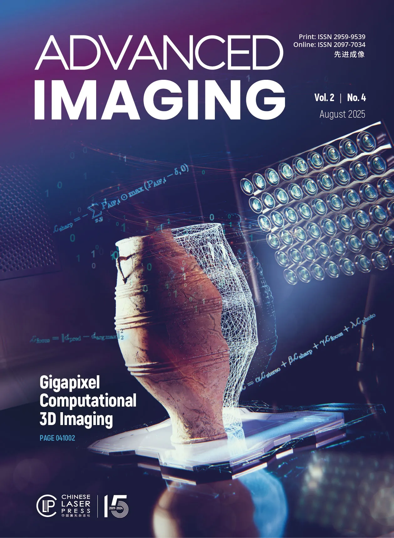
We present a novel approach for capturing gigapixel-resolution micron-scale three-dimensional (3D) images of large complex macroscopic objects using a multi-camera array paired with a custom 3D reconstruction algorithm. Our system overcomes inherent trade-offs among resolution, field of view (FOV), and depth of field (DOF) by capturing stereoscopic focal stacks across multiple perspectives, enabling an effective FOV of approximately degrees and capturing surface depth maps at lateral resolutions less than 40 µm and depth resolutions of about 0.5 mm. To achieve all-in-focus RGB (red, green, and blue) composites with precise depth, we employ a novel self-supervised neural network that integrates focus and stereo cues, resulting in highly accurate 3D reconstructions robust to variations in lighting and surface reflectance. We validate the proposed approach by scanning 3D objects, including those with known 3D geometries, and demonstrate sub-millimeter depth accuracy across a variety of scanned objects. This represents a powerful tool for digitizing large complex forms, allowing for near-microscopic details in both depth mapping and high-resolution image reconstruction.
We present the Fourier lightfield multiview stereoscope (FiLM-Scope). This imaging device combines concepts from Fourier lightfield microscopy and multiview stereo imaging to capture high-resolution 3D videos over large fields of view. The FiLM-Scope optical hardware consists of a multicamera array, with 48 individual microcameras, placed behind a high-throughput primary lens. This allows the FiLM-Scope to simultaneously capture 48 unique 12.8 megapixel images of a 28×37mm field-of-view, from unique angular perspectives over a 21deg×29deg range, with down to 22μm lateral resolution. We also describe a self-supervised algorithm to reconstruct 3D height maps from these images. Our approach demonstrates height accuracy down to 11μm. To showcase the utility of our system, we perform tool tracking over the surface of an ex vivo rat skull and visualize the 3D deformation in stretching human skin, with videos captured at up to 100 frames per second. The FiLM-Scope has the potential to improve 3D visualization in a range of microsurgical settings.
Convulsive seizure behaviors are a hallmark feature of epilepsy, but automated detection of these events in freely moving animals is difficult. Here, we employed the Ramona Kestrel™, a high-resolution multi-camera array microscope with high-speed video acquisition and custom supervised machine learning (ML) for automated detection of larval zebrafish between 3- and 7-days post-fertilization (dpf). We assessed data from over 2700 zebrafish either exposed to a chemoconvulsant (pentylenetetrazole, PTZ) or genetic zebrafish lines representing Developmental Epileptic Encephalopathy (DEE) syndromes.
This paper presents a validation of the Ramona Kestrel™ for automated, high-throughput screening for early-stage drug development and toxicology assessment with zebrafish embryos. The Kestrel™ is a novel high-throughput imaging platform featuring a 24-camera array that enables simultaneous acquisition of high-resolution video data across 96-well plates. This paper validated the system using zebrafish embryonic photomotor response (EPR) assays, demonstrating its ability to track behavioral responses in chorionated and dechorionated embryos without workflow modifications. The system successfully detected concentration-dependent responses to ethanol, methanol, and bisphenol A across different plate formats and well volumes
Here, we present reflective Fourier light field computed tomography (ReFLeCT), a high-speed volumetric fluorescence computational imaging technique. ReFLeCT synchronously captures entire tomograms of multiple unrestrained, unanesthetized model organisms across multi-millimeter 3D FOVs at 120 volumes per second. In particular, we applied ReFLeCT to reconstruct 4D videos of fluorescently labeled zebrafish and Drosophila larvae, enabling us to study their heartbeat, fin and tail motion, gaze, jaw motion, and muscle contractions with nearly isotropic 3D resolution while they are freely moving.
Patient-derived organoids (PDOs) are a valuable tool for investigations of intra-tumor and inter-site heterogeneity and patient-specific drug responsiveness. In this work, we employ the MCAM (Multi-Camera Array Microscope) Vireo™ system to rapidly acquire brightfield images of PDOs derived from gastrointestinal surgical resection samples in under 2 minutes per 24 well plate and a new machine learning model to automatically analyze this data.
We present a multi-camera array for capturing dynamic high-resolution videos of the human face. Compared to traditional single-camera configurations, our array of 54 individual cameras allows stitching of high-resolution composite video frames (709 megapixels total). In our novel multi-focus strategy, each camera in the array focuses on a unique object plane to resolve non-planar surfaces at a higher resolution than a standard single-lens camera design. Compared to a single-focus configuration, this is almost a 10-fold increase in effective DOF. We demonstrate how our multi-focus camera array can capture dynamic facial expressions at microscopic resolution with relevance in several biomedical applications.
We applied the Ramona Vireo to rapidly (< 1 min.) capture and process 3D image data of up to 96 cortical organoids grown within multi-well plates. This study aimed to longitudinally measure and analyze the growth of PGP1 line iPSC-derived cortical organoids at high throughput to evaluate the impact of morphogens (valproic acid and lithium) on cortical organoid growth. Results revealed a dose-dependentdecrease in organoid area with increasing concentrations of valproic acid, whereas lithium induced a moderate yet consistent increase. These findings underscore the power of high-throughput imaging andsegmentation for quantifying how drugs and dosages influence cortical organoid growth in vitro.
Here, we describe a cell viability assay and workflow using the Vireo by Ramona that offers a 10-fold decrease in processing time compared to traditional imaging-based viability assessments. It also offers a more accurate workflow compared to standard CellTiter-Glo (CTG) assays. This technology can revolutionize the field of drug discovery and toxicology by drastically decreasing the time between image acquisition and data analysis and allowing for high-throughput screening capabilities.
We demonstrate how the Ramona Kestrel can be used to capture information about zebrafish (D. rerio) larvae at various spatial and temporal scales from 1 to 5 days post fertilization. At these stages, the zebrafish larvae can be placed in 96 well plates where they be exposed to different chemicals in order to study their toxicological effects at high throughput prior to conducting studies in other animal models or humans.
Clinical diagnosis of cytology specimens is especially challenging given that samples are both spread over large areas and thick, which requires 3D capture. Here, we introduce a new parallelized microscope for scanning thick specimens across extremely wide fields-of-view (54 × 72 mm2) at 1.2 and 0.6 μm resolutions, accompanied by machine learning software to rapidly assess these 16 gigapixel scans. This Multi-Camera Array Scanner (MCAS) comprises 48 micro-cameras closely arranged to simultaneously image different areas. By capturing 624 megapixels per snapshot, the MCAS is significantly faster than most conventional whole-slide scanners.
We present a large-scale computational 3D topographic microscope that enables 6-gigapixel profilometric 3D imaging at micron-scale resolution across >110 cm2 areas over multi-millimeter axial ranges. We developed a self-supervised neural network-based algorithm for 3D reconstruction and stitching that jointly estimates an all-in-focus photometric composite and 3D height map across the entire field of view, using multi-view stereo information and image sharpness as a focal metric. Validation experiments on gauge blocks demonstrate a profilometric precision and accuracy of 10 µm or better, with subsequent experiments demonstrating the broad utility of our new computational microscope in applications ranging from cultural heritage to industrial inspection.
This study introduces a rapid, high-throughput approach using the Multi-Camera Array Microscope (MCAM™) to image and quantify neutrophils in zebrafish embryos—a key indicator of environmental impacts on immune function. Using advanced machine learning, the system is able to process and count individual fluorescent neutrophils across a 96-well plate in just over 5 minutes, offering a rapid and accurate alternative to traditional manual counting methods.
This article demonstrates how wide-field-of-view microscopy with Ramona’s Multi-Camera Array Microscope (MCAM™) can resolve three-dimensional information at high speed and spatial resolution. It then shows how this technology can serve as a powerful tool for studying the behavior of freely moving organisms, such as ants, fruit flies, and zebrafish larvae.
This publication presents results from three unique Multi-Camera Array Microscope (MCAM™) configurations for different use cases. These configurations include simultaneous capture with 3D object depth estimation, continuous video capture at high resolution over a large field of view, and a high-resolution configuration to produce 9.8 GP composites of large histopathology specimens.
This paper details how Ramona’s Multi-Camera Array Microscope (MCAM™) enables comprehensive high-resolution recording from multiple spatial scales simultaneously, ranging from cellular-scale structures to large-group behavioral dynamics. This allows researchers to observe the behavior and fine anatomical features of numerous freely moving model organisms on multiple spatial scales, including larval zebrafish, fruit flies, nematodes, carpenter ants, and slime mold.
This research demonstrates a machine learning technique for swift and precise insect egg identification using Ramona's Multi-Camera Array Microscope (MCAM™), distinguishing two crucial pest species with more than 99% accuracy. Validated with around 5500 images, the approach suggests new avenues for real-time agricultural pest diagnostics.
This paper details how Ramona’s Multi-Camera Array Microscope (MCAM™) can quantify morphological features in bacterial colonies across multi-well plates. It shows how the system can be used to augment high-throughput assays by synchronously capturing valuable phenotypic information throughout an acquisition and analysis pipeline.


.avif)